
Anatomy of the Foot and Ankle OrthoPaedia
These bones are arranged in two rows, proximal and distal. The bones in the proximal row form the hindfoot, while those in the distal row from the midfoot. Hindfoot. Talus. Calcaneus. The talus connects the foot to the rest of the leg and body through articulations with the tibia and fibula, the two long bones in the lower leg. Midfoot. Navicular.

Foot Bone Anatomy Vector Illustration 539973 Vector Art at Vecteezy
Use these bones of the foot quizzes to master your identification skills. Overview of the bones of the foot and their divisions into the hindfoot, midfoot and forefoot. With a total of 26 bones in each foot, learning the bony anatomy of the foot is no piece of cake. That is, the memorization aspect.

Foot Description, Drawings, Bones, & Facts Britannica
The navicular bone is found on the inner side of the foot. The navicular articulates with five of the other tarsal bones - at the top with the talus, talonavicular joint, laterally (outer side) with the cuboid, cubonavicular joint, and at the bottom it articulates with the three cuneiform bones. In around 10% of the population, a small extra piece of bone develops next to the navicular which.

How to have beautiful, healthy feet banish bunions and other abominations! Harrogate Yoga
The foot is a part of vertebrate anatomy which serves the purpose of supporting the animal's weight and allowing for locomotion on land. In humans, the foot is one of the most complex structures in the body. It is made up of over 100 moving parts - bones, muscles, tendons, and ligaments designed to allow the foot to balance the body's.
.jpg)
Foot Bone Diagram resource Imageshare
The 26 bones of the foot consist of eight distinct types, including the tarsals, metatarsals, phalanges, cuneiforms, talus, navicular, and cuboid bones. The skeletal structure of the foot is.

Foot & Ankle Bones
The foot is a complex structure comprised of over 26 bones, 30 joints, numerous tendons, ligaments, and muscles responsible for our ability to stand upright, supporting the weight of the entire body and provides the base for the mechanism for bipedal gait. The foot corresponds to the portion of the lower extremity distal to the ankle and divides into hind, mid and forefoot.

Foot & Ankle Bones
The foot is the region of the body distal to the leg and consists of 28 bones. These bones are arranged into longitudinal and transverse arches with the support of various muscles and ligaments. There are three arches in the foot, which are referred to as: Medial longitudinal arch. Lateral longitudinal arch.
.jpg)
Foot Bone Diagram resource Imageshare
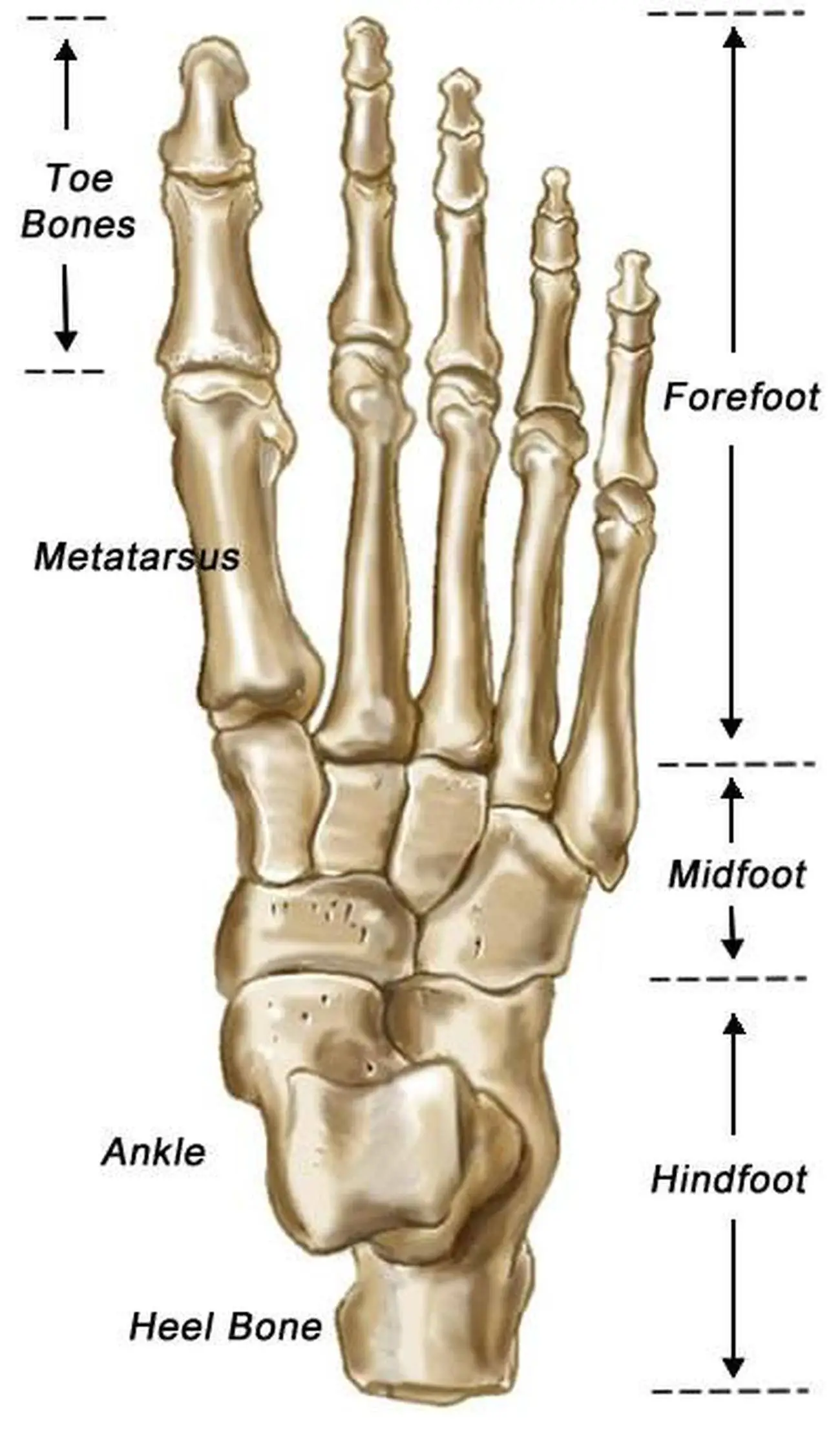
Foot Bones: Forefoot. The forefoot consists of 19 bones; 5 metatarsal bones and 14 phalanges. The big toe has 2 phalanges bones, while the remaining four have 3 phalanges each. The 1st metatarsal is the shortest and thickest of the metatarsals, and it is designed to take up to 40% of your body weight in standing, which rises to 70% when walking.

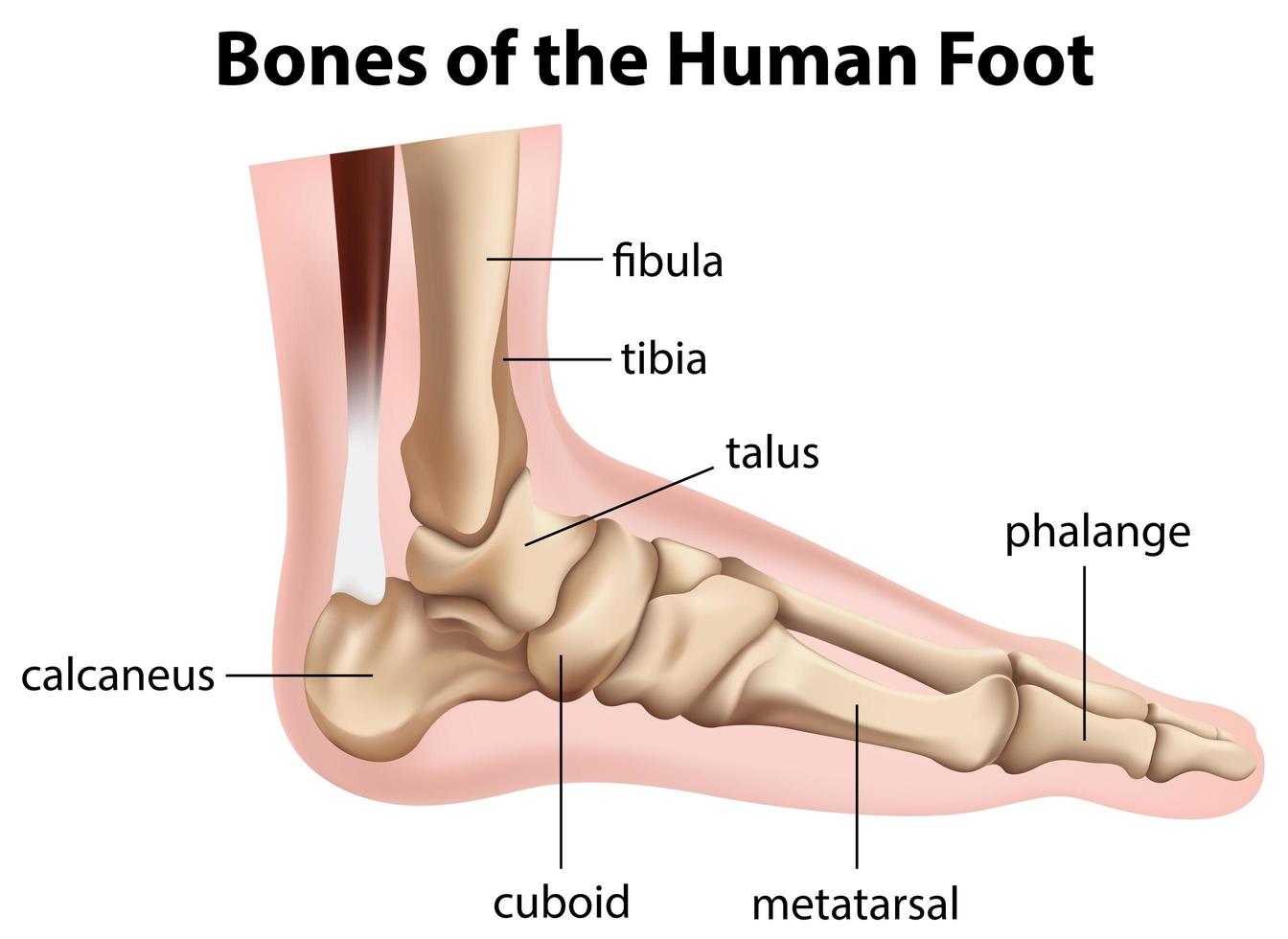
Foot bones Anatomy, conditions, and more (2022)
Tarsals. The tarsals are a group of seven bones close to the ankle. The proximal tarsal bones are the talus and the calcaneus, which is the largest bone of the foot. The talus is on top of the.

Foot skeleton composed of 28 skeletal bones. It can be divided into... Download Scientific Diagram
Fore-foot - the fore-foot is composed of the metatarsals and phalanges. The bones that comprise the fore-foot are those that are last to leave the ground during walking. Mobile Joints of the foot and ankle: (See Figure 3.) Ankle joint. Sub-talar joint. Talo-navicular joint. Metatarso-phalangeal (MTP) joints.
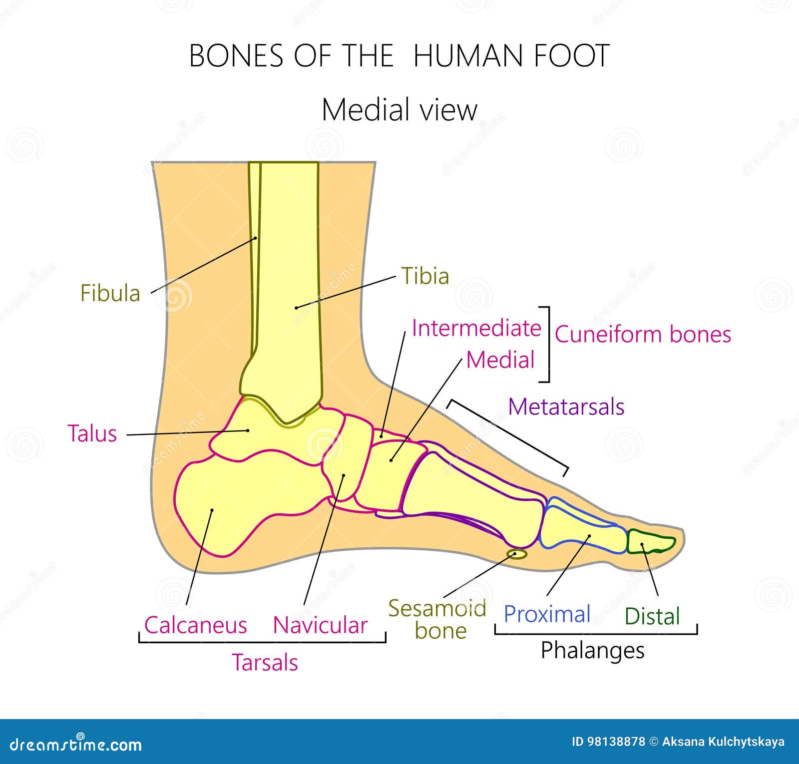
Anatomy_bones of the Human Foot Medial View Stock Vector Illustration of fibula, phalanx 98138878
A solid understanding of anatomy is essential to effectively diagnose and treat patients with foot and ankle problems. Anatomy is a road map. Most structures in the foot are fairly superficial and can be easily palpated. Anatomical structures (tendons, bones, joints, etc) tend to hurt exactly where they are injured or inflamed.

Foot Description, Drawings, Bones, & Facts Britannica
The bones of the foot are organized into rows named tarsal bones, metatarsal bones, and phalanges. These make up the toes and broad section of the feet. The other bones of the foot that create the.
Bones of the human foot diagram 1142236 Vector Art at Vecteezy
Ankle anatomy. The ankle joint, also known as the talocrural joint, allows dorsiflexion and plantar flexion of the foot. It is made up of three joints: upper ankle joint (tibiotarsal), talocalcaneonavicular, and subtalar joints.The last two together are called the lower ankle joint. The upper ankle joint is formed by the inferior surfaces of tibia and fibula, and the superior surface of talus.

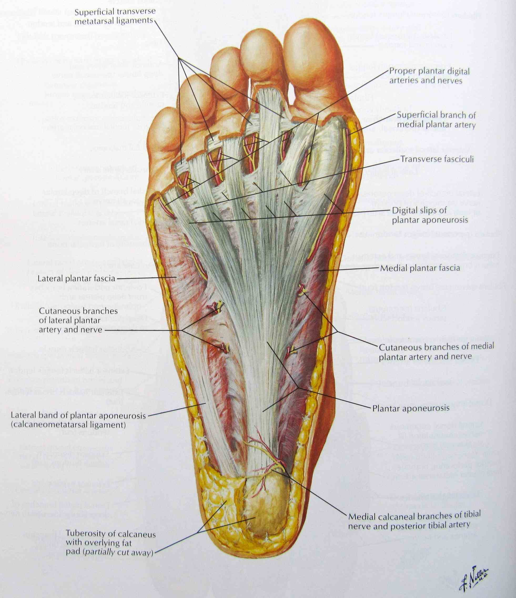
The bones in the foot inferior view (Picture illustrated from Thieme... Download Scientific
Calcaneus: The largest bone of the foot, it is commonly referred to as the heel of the foot. It points upward, while the remaining bones of the feet point downward. Talus: This irregularly shaped.
Anatomy The Bones Of The Foot
The bones of the foot provide mechanical support for the soft tissues; helping the foot withstand the weight of the body whilst standing and in motion.. They can be divided into three groups: Tarsals - a set of seven irregularly shaped bones.They are situated proximally in the foot in the ankle area. Metatarsals - connect the phalanges to the tarsals.
Pictures Of Bones Of The Feet
Summary. The foot is an intricate part of the body, consisting of 26 bones, 33 joints, 107 ligaments, and 19 muscles. Scientists group the bones of the foot into the phalanges, tarsal bones, and.