
auriculair diagram Google zoeken Ooracupunctuur Pinterest
1 Recommendation Popular answers (1) yes Adil is right you can refer to these definitions The left auricle, also known as the left atrial appendage (LAA), is actually a small, muscular pouch.

Difference Between Atrium and Auricle Definition, Anatomy, Physiology
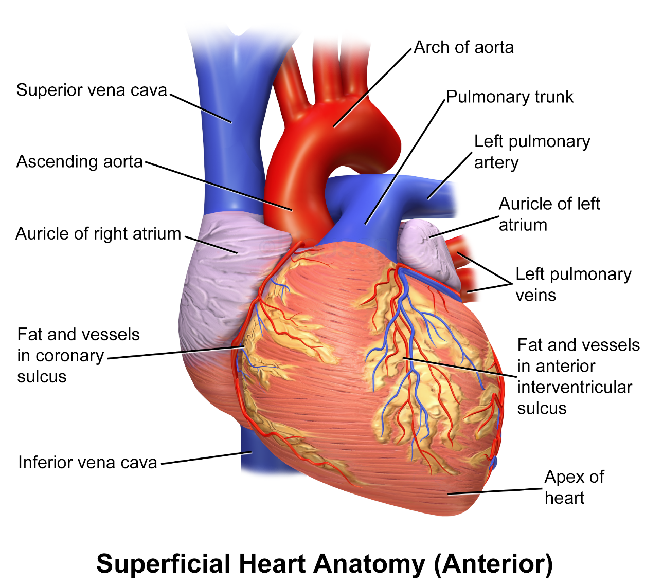
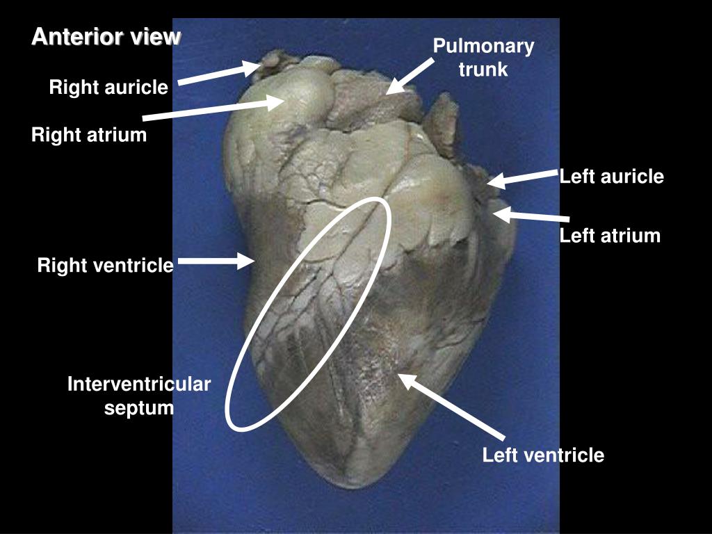
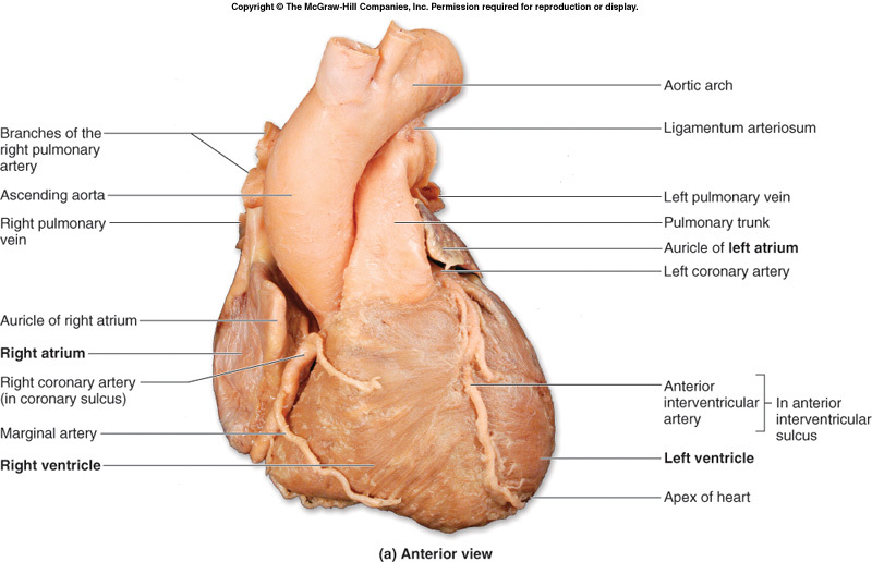
The Right Auricle By: Tim Taylor Last Updated: Jun 11, 2015 Anatomy Explorer Heart Aortic Valve Bundle Branches Chordae Tendineae Interventricular Septum Left Atrium Left Auricle Left Ventricle Mitral Valve Papillary Muscles Pulmonary Valve Purkinje Fibers Right Atrium Right Auricle Right Ventricle Tricuspid Valve Pulmonary Trunk

The Auricles of the Heart Left and Right YouTube
Auricles are relatively thin-walled structures that can fill with blood and empty into the atria or upper chambers of the heart. You may also hear them referred to as atrial appendages.. Although the ventricles on the right and left sides pump the same amount of blood per contraction, the muscle of the left ventricle is much thicker and.
Describe the internal structure of the human heart.
The Surprising Differences Between the Right and Left Auricles of the Heart.

PPT Chapter 18 Anatomy of the Cardiovascular System PowerPoint
The left and right auricles play a crucial role in maintaining a healthy heart. They help to regulate blood flow and ensure that the heart is pumping efficiently. If the auricles are not functioning correctly, it can lead to a range of heart problems, including: Arrhythmias Atrial fibrillation Heart failure Stroke How Can Nao Medical Help?
PPT Sheep Heart Dissection PowerPoint Presentation, free download
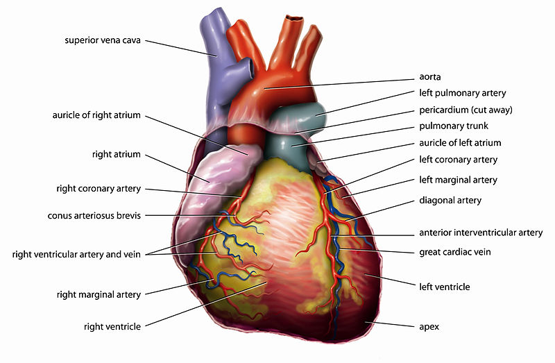
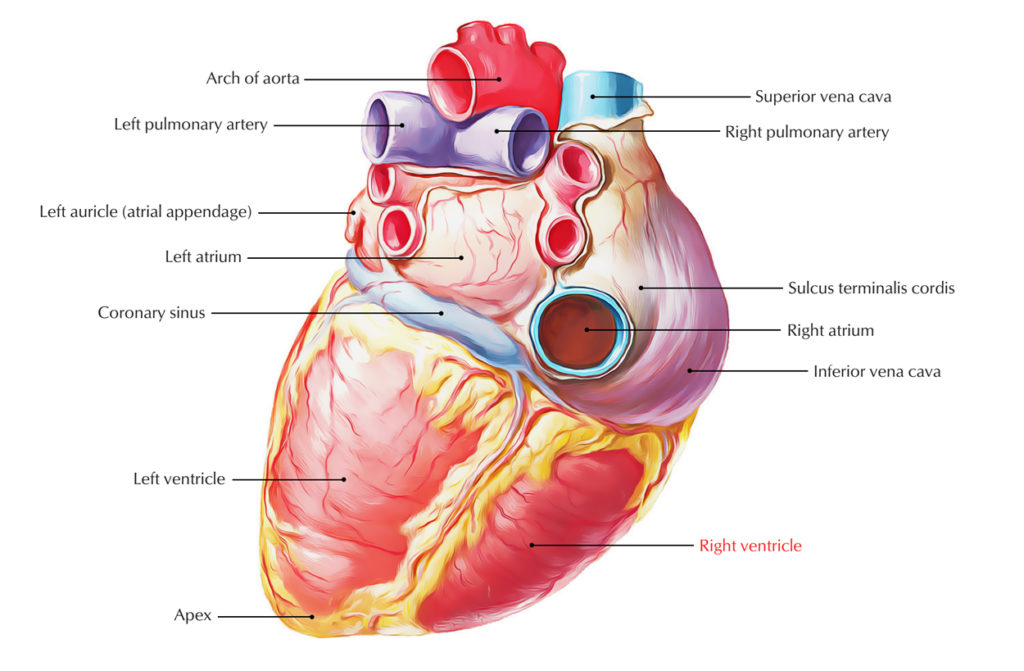
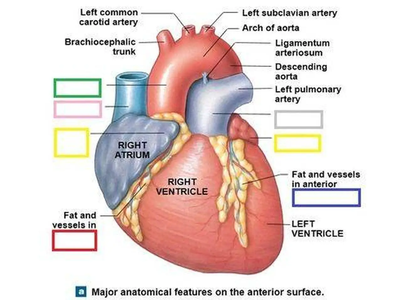
The right margin is the small section of the right atrium that extends between the superior and inferior vena cava . The left margin is formed by the left ventricle and left auricle. The superior margin in the anterior view is formed by both atria and their auricles. The Inferior margin is marked by the right ventricle.

PPT The Circulatory System The Heart PowerPoint Presentation, free
Right Auricle Right Brachiocephalic Vein
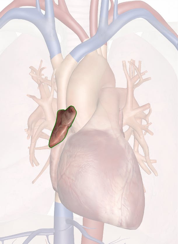
The Right Auricle Anatomy and 3D Illustrations
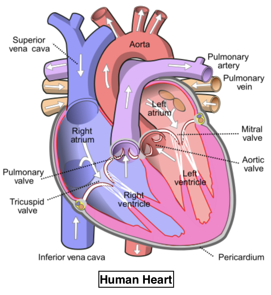
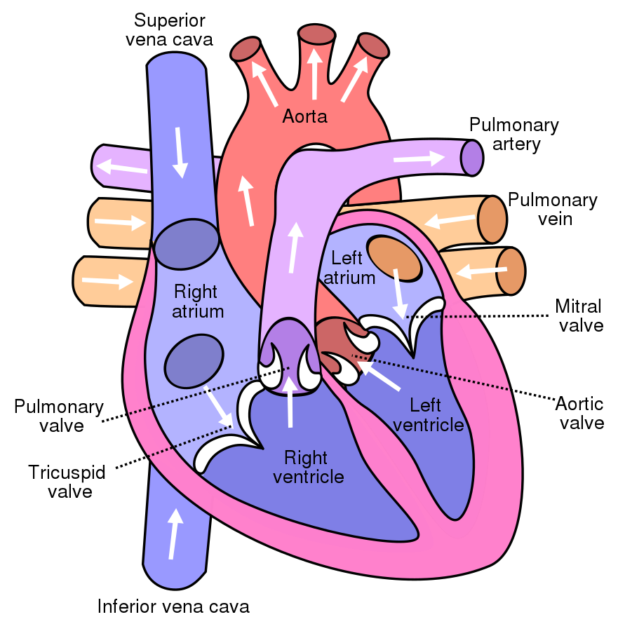
The heart is divided into 4 chambers: 2 upper chambers for receiving blood from the great vessels, known as the right and left atria, and 2 stronger lower chambers, known as the right and left ventricles, which pump blood throughout the body.

HUMAN BIOLOGY AND HEALTH CIRCULATION
The auricles of the right and left atrium are prominent anatomic structures of the heart. The anatomic area of the right atrial auricle, which lies in the proximity of the basic rhythm center of the heart, the sinus node, is used for the venous cannulation of the heart before the extracorporeal circulation is entered.
c. Circulatory System BIOLOGY4ISC
The bulbus cordis develops into the right ventricle. The primitive ventricle forms the left ventricle. The primitive atrium becomes the anterior portions of both the right and left atria, and the two auricles. The sinus venosus develops into the posterior portion of the right atrium, the SA node, and the coronary sinus.
Difference Between Auricle and Ventricle Definition, Structure
Extending from the antero-medial portion of the chamber is the right auricle (right atrial appendage) - a muscular pouch that acts to increase the capacity of the atrium. The interior surface of the right atrium can be divided into two parts, each with a distinct embryological origin.
The valve present between the left auricle and left ventricle is (a
In Fig. 1 the bifurcation of the main bundle into right and left septal divisions is shown; in Fig. 2 is depicted the distribution of the left septal division within the left ventricle of the calf's heart. The branch appears on the septal wall of the ventricle about two centimetres below the aortic orifice; just below the aortic orifice the bundle is buried beneath a thick layer of muscle (cut.
The lower chambers of the heart are calleda. Auriclesb. Ventriclesc
an imaginary line passing through the jugular notch superiorly, a parallel line passing through the xiphoid process inferiorly, bilaterally are two imaginary vertical lines, each passing through the respective left and right midclavicular lines (or nipple line in the non-pendulous breast ).
Right Ventricle Earth's Lab
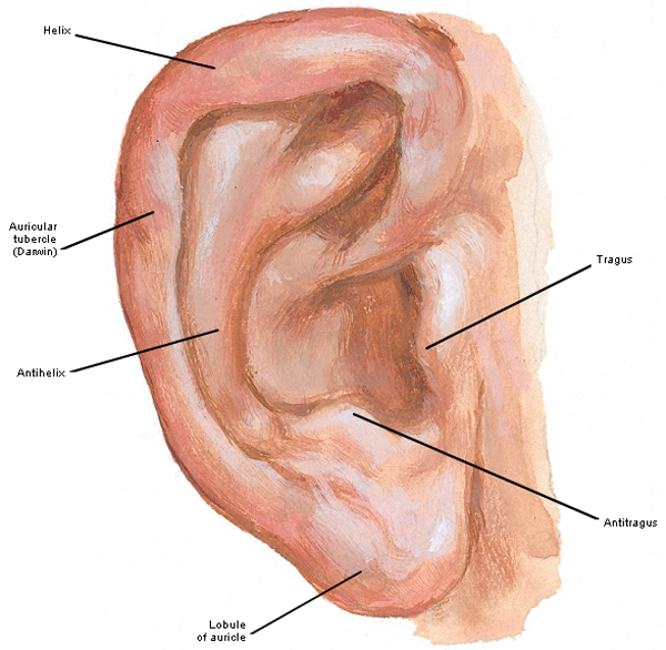
An auricle consists of skin over contoured cartilage, and it is held in place by muscles and ligaments. Shape may differ by body type and person. Auricles are located on both sides of the head.
“Hear, Here The Ear”
Auricles, also known as atria, are the two uppermost blood-receiving chambers of the heart that receives the blood from the veins. These are smaller and thin-walled as compared to the ventricles as they don't need to generate the force of contraction to pump the blood out of the heart. There are two atria, the right, and the left atrium.
Pictures Of Atrial Appendage (auricle)
The auricle or auricula is the visible part of the ear that is outside the head. It is also called the pinna ( Latin for ' wing ' or ' fin ', pl.: pinnae ), a term that is used more in zoology . Structure The diagram shows the shape and location of most of these components: antihelix forms a 'Y' shape where the upper parts are: